Our state-of-the-art center is a fully-equipped facility where we identify the nature of an illness or other problems using a combination of accurate examination and advanced technology.
Vision Care Center is equipped with the world’s most advanced diagnostic equipment. Our expert and experienced team of doctors are capable of accurately interpreting the diagnostic reports and results.
With the help of our diagnostic equipment, we can conduct the following tests:
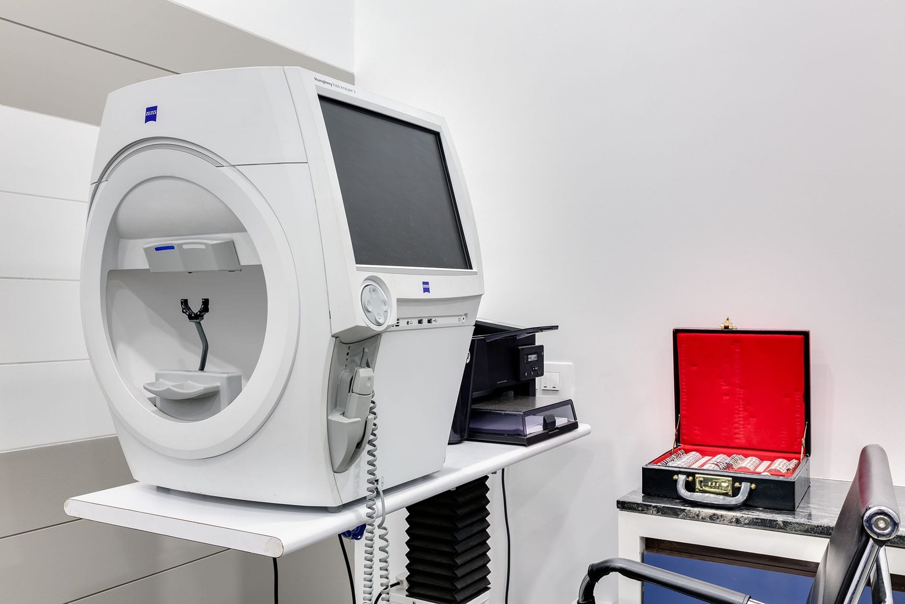
Humphreys Visual Field HFA-III 840 Zeiss
A visual field test is an eye examination that can detect dysfunction in central and peripheral vision, which may be caused by various medical conditions such as glaucoma, stroke, pituitary disease, brain tumours or other neurological deficits. Visual field testing is performed clinically by keeping the subject's gaze fixed while presenting lights of different intensity at various places within their visual field.
Vision Care Center is equipped with the world’s most advanced ZEISS Humphrey Field Analyzer 3 that reduces the testing time as well as gives more insight into the glaucoma management.
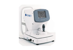
Corneal Topography
This is a non-invasive medical imaging technique to map the surface curvature of the cornea, the outer structure of the eye. Since the cornea is normally responsible for about 70% of the eye's refractive power, its topography is of critical importance in determining the quality of vision and corneal health. .
The topography map is, therefore, a valuable aid to the examining ophthalmologist or optometrist and can assist in the diagnosis and treatment of a number of conditions like Keratoconus. It helps to plan cataract surgery and intraocular lens.
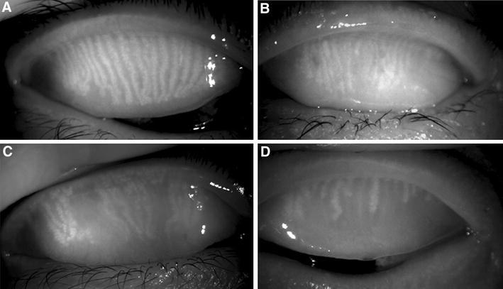
Meibography
Meibography is an imaging study developed exclusively for the purpose of observing the morphology of meibomian glands. It is the specialized technique of visualizing meibomian glands in the eyelids of the eye with the help of infrared light-based technology. Meibomian glands play a significant role in tear production by contributing lipids to the superficial tear film. Dysfunction of the meibomian glands destabilizes tears resulting in evaporative dry eye. Therefore, it is the major cause of severe dry eye and results in a decrease in quality of life.
Vision Care Center is equipped with Meibography technology to help us understand your dry eye disease better and treat it effectively.
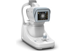
Non-Contact Tonometry
This test is done to evaluate the pressure inside the eye without touching the organ. This technology helps us to measure the intraocular pressure with the help of air puff. It helps in the management of glaucoma - a disease that damages the optic nerve of the eye, usually resulting in vision loss or even blindness.
We use a tool called a tonometer that blows a tiny puff of air, measuring eye pressure indirectly by the eye's resistance to the puff.
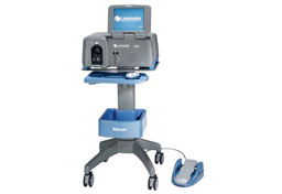
Alcon Laureate World Phacoemulsification System
We use this system to perform sutureless cataract surgeries effectively. This cutting edge phacoemulsification technology allows surgeons more control in the procedure than they have ever had before. Followability is increased, and some of the known hazards of phaco, especially thermal injury to the eye, are reduced. By using this latest phacoemulsification system, our surgeons are able to cure any grade cataract.
Class B Autoclave Hanshin ( CE certified)
We use this to make sure instruments being used in the eye are completely sterilized. We understand the importance of eyes and that any small infection can snowball into infectious disease. That's why, we ensure all our instruments, which are used in surgery, are thoroughly sterilised with this lastest steam sterilizer device.
This typical B-Class small steam sterilizer is optimum to sterilize the medical devices including a narrow and long lumen(hollow loads) used frequently in our hospital, and all the general medical supplies effectively.
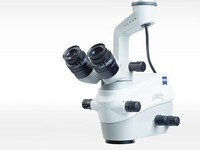
Zeiss Operating Microscope
We use this to visualize finer details of your eye while doing cataract surgery. It is important for surgeons to visualize and analyse the finer details of the eye while doing surgery. ZEISS ophthalmic microscope provides excellent visualization of the eye, with optimal ergonomics and integrated assistance functions.
This advanced operating microscope helps us to actively track and analyze the finer details when performing the procedure, ensuring safety and results in a successful procedure.
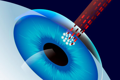
Collagen Cross-Linking System
Keratoconus is the disease of the cornea where the cornea becomes thin and starts protruding from the surface. Collagen cross-linking makes the cornea stiff and stabilizes the corneal structure.
Corneal collagen cross-linking is a technique which uses UV light and a photosensitizer to strengthen chemical bonds in the cornea. The goal of the treatment, with the help of this system, is to halt progressive and irregular changes in the corneal shape known as ectasia. Because ectasia can often lead to high levels of myopia and astigmatism.
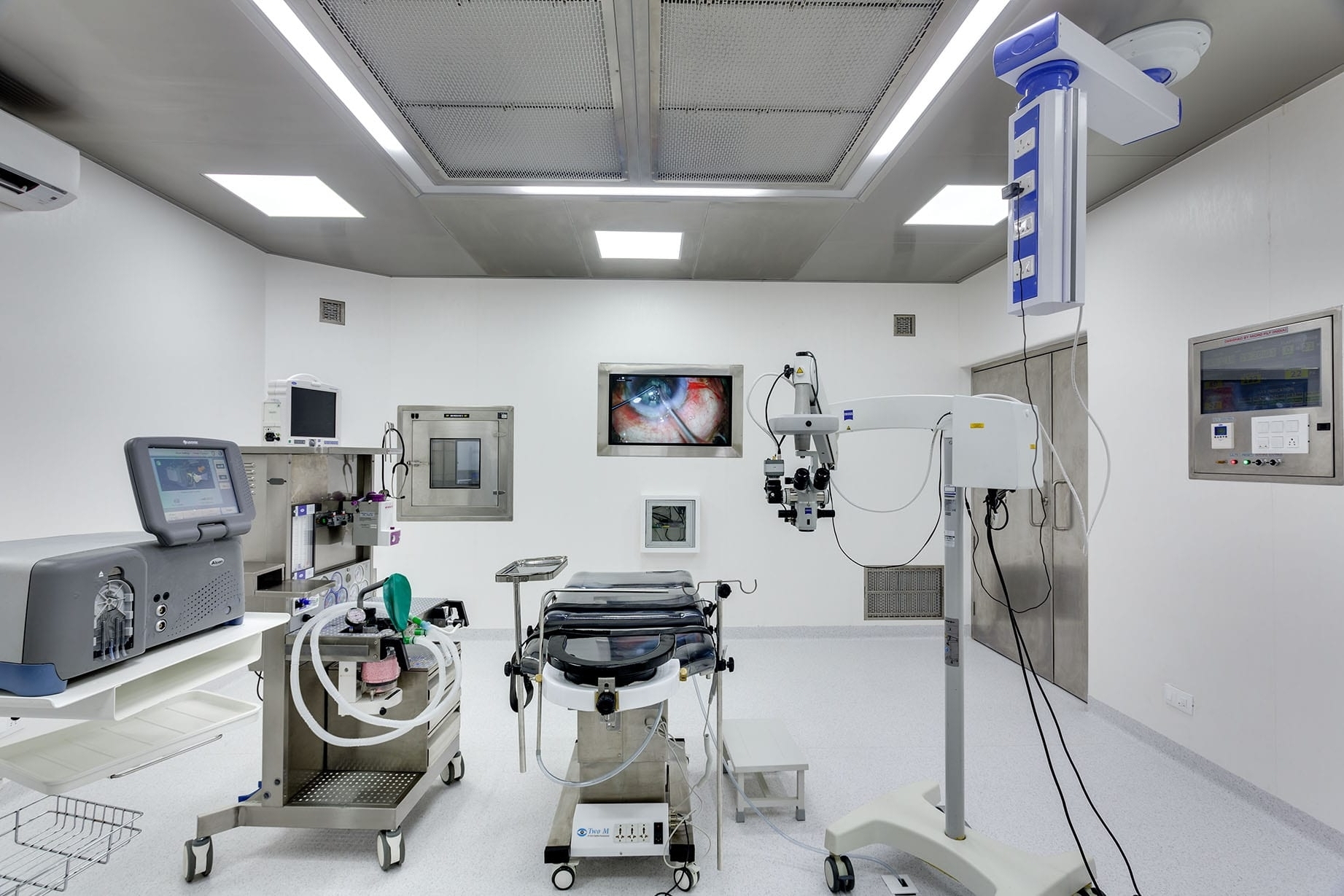
Advanced Modular Operation Theatre
For doing any surgery, a clean environment is an absolute necessity. Our modular operation theatre has walls and floor made up of Polyvinyl chloride, which prevents the growth of any organism on the surface. The modular OT also has an air handling unit with HEPA filters, which ensures clean air supply all the time, to make your eye surgery safe and prevent infection.
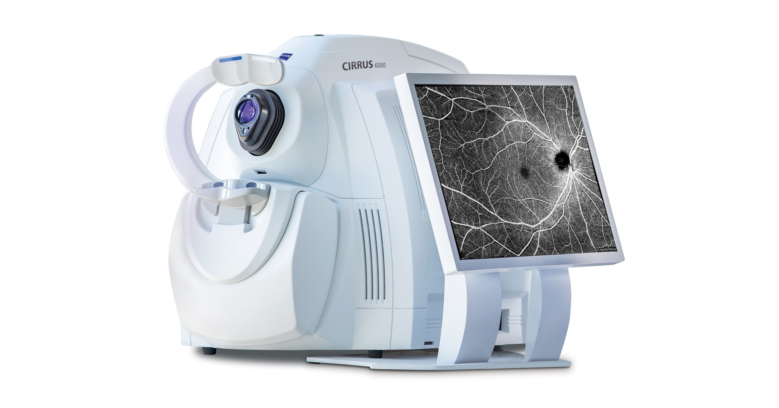
ZEISS CIRRUS HD OPTICAL COHERENCE TOMOGRAPHY (OCT)
Optical Coherence Tomography (OCT) is a non-invasive imaging test. OCT uses light waves to take cross-section pictures of your retina. With OCT, your doctor can see each of the retina’s distinctive layers. This allows them to map and measure their thickness. These measurements help with the diagnosis of diseases related to retina and glaucoma and guide the treatment.
We are equipped with Revo Optopol Optical Coherence Tomography, which is the world’s most advanced OCT and works on the principle of spectral domain. With this, our doctors are confident about treating retina-related and glaucoma diseases.
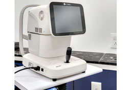
OA-2000 Optical Biometry
Biometry of the eye involves taking measurements of the eye, including the Axial Length (AL), Keratometry and Anterior Chamber Depth (ACD). These measurements are crucial for the selection of the correct lens power in order to achieve the minimal spectacle number after cataract surgery. Therefore, ocular biometry is an essential step before cataract surgery. There are currently two procedures available: Ultrasound and Optical Biometry.
Vision Care Center is equipped with advanced Optical Biometry, which works on the principle of the Fourier domain Axial Length measurement and topography as well as ultrasound biometry. As a result, we can offer customized intraocular lens based on most accurate measurements to keep you spectacle-independent.
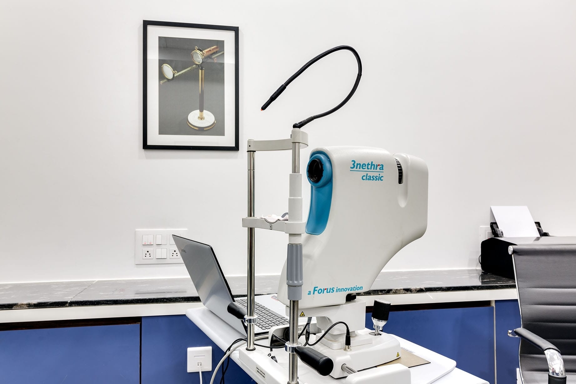
Fundus Photography
Fundus photography involves photographing the retina and optic disc of the eye, which is also known as the fundus. The main purpose of photographing it is to document the disease process, which helps in diagnosing the progression of the problem and the treatment required.
Vision Care Center is equipped with an advanced fundus photography system.
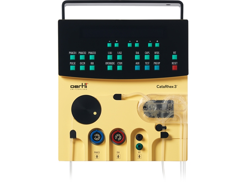
MIGS HFDS FOR GLUACOMA
Swiss-made Oertli provides surgical treatment of glaucoma as micro‑invasive glaucoma surgery (MIGS). High-Frequency Deep Sclerotomy (HFDS) creates a direct access between the anterior chamber and the Schlemm canal that bypasses the outflow resistance of the trabecular meshwork. We are the very few hospitals in India offering this treatment modality to our glaucoma patients.